Drawing Of Gallbladder
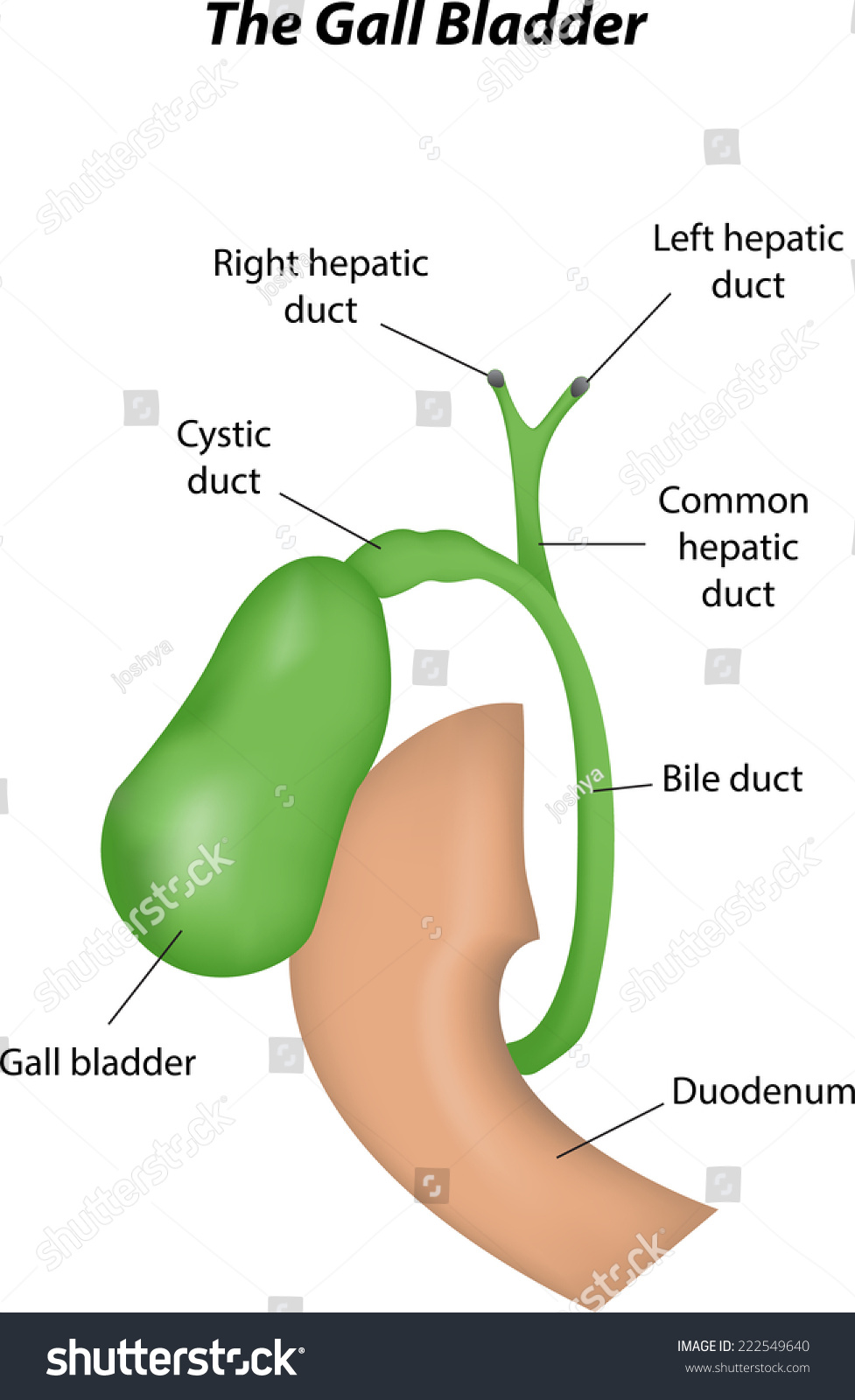
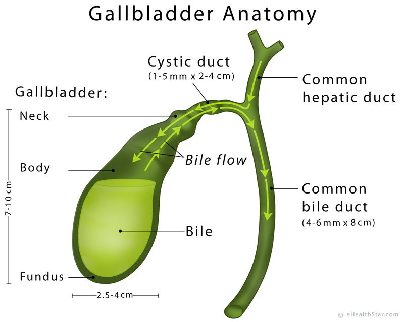
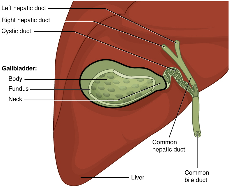
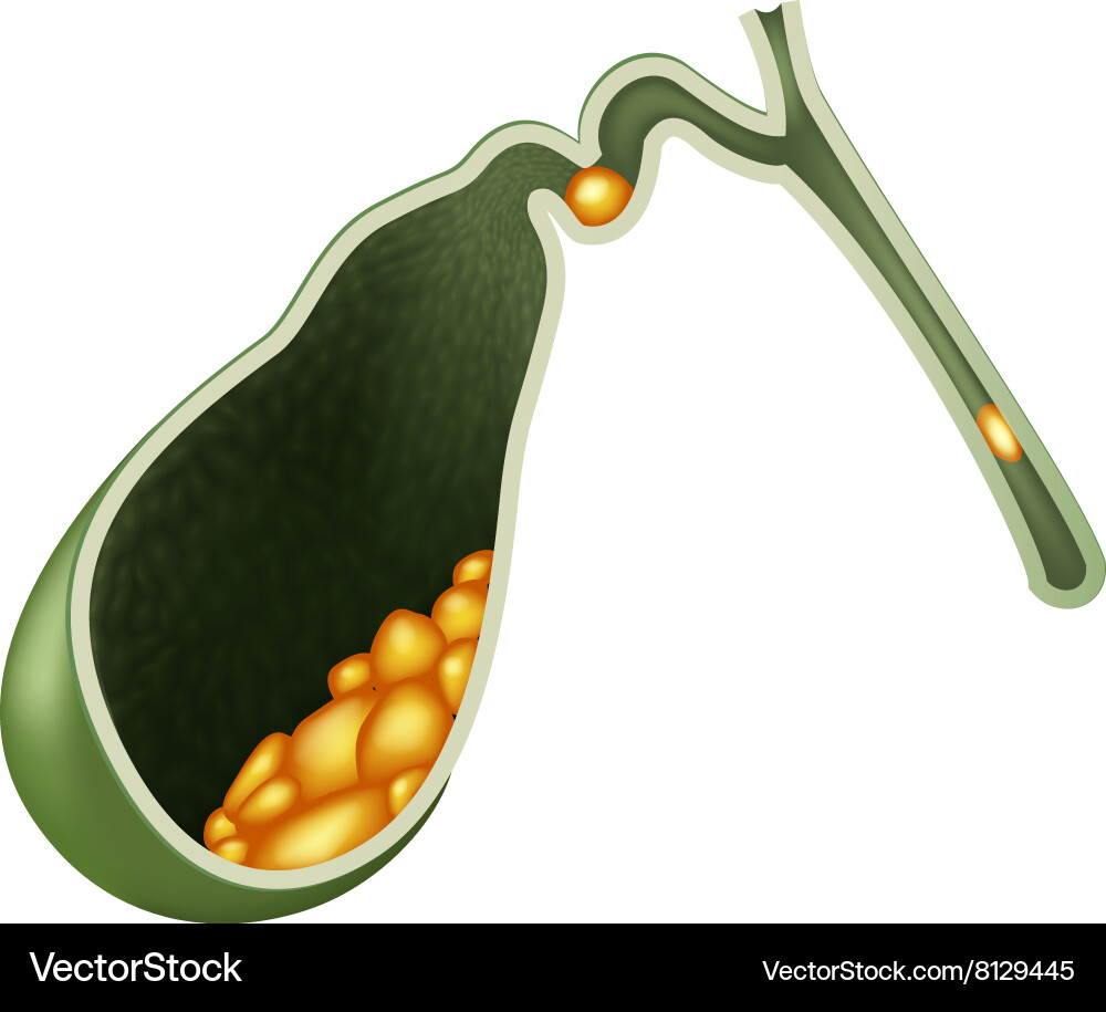
Drawing Of Gallbladder - A gallbladder scan is a type of nuclear. Web this will teach you how to draw gallbladder and name the parts. This muscular organ also concentrates and releases bile into the digestive system. Web the gallbladder is a hollow organ that sits beneath the liver and stores bile made in the liver. It’s a small organ, shaped like a pear, that holds a fluid called bile. The biliary system is a series of ducts within the liver, gallbladder, and pancreas that empty into the small intestine. Web as part of the gustatory response, the stored bile is then released from the gallbladder in response to cholecystokinin. You can locate the gallbladder between the lateral aspect of the rectus abdominis muscle and the right costal margins. Your gallbladder sits on the right side of your belly, below your liver. The next layer is a layer of loose connective tissue called the lamina propria. Structurally it is characterized by its pyriform shape and location between the. The liver produces and releases bile, a green substance that absorbs and breaks down lipids. Your gallbladder sits on the right side of your belly, below your liver. The inside of the gallbladder is comprised of columnar epithelium cells. The neck connects to a system of ducts. This article will review the anatomy of the gallbladder as well as the associated biliary apparatus. The most common issue you may develop with your gallbladder is gallstones. The bile duct is drained by the superior pancreaticoduodenal vein, which in turn empties into the hepatic portal system. Hello friends, this is my youtube. Also, press the bell icon to get. Web this will teach you how to draw gallbladder and name the parts. Drawing of the biliary system, with the liver, gallbladder, duodenum, pancreatic duct, common bile duct, pancreas, cystic duct, and hepatic ducts. Your gallbladder stores and releases bile to help your digestive system break down fats. Special attention will also be paid to the relevant embryology, histology and. This muscular organ also concentrates and releases bile into the digestive system. Your gallbladder stores and releases bile to help your digestive system break down fats. Web the gallbladder is a hollow organ that sits beneath the liver and stores bile that is made in the liver. This article will review the anatomy of the gallbladder as well as the. Findings in this randomized clinical trial of 90 participants, both gc and ccrt. Your gallbladder stores and releases bile to help your digestive system break down fats. Please don’t forget to subscribe to my video. The gallbladder is a small hollow organ about the size and shape of a pear. Test your knowledge on the gallbladder with this quiz. This liquid, made in your. Web what it does. When bile is needed, the gallbladder contracts, forcing the fluid through a tube called the cystic duct. Question do the regimens of adjuvant gemcitabine and cisplatin (gc) and capecitabine concurrent with chemoradiation (ccrt) with capecitabine result in adequate early survival rates in patients with resected stage ii and stage iii gallbladder. Web this will teach you how to draw gallbladder and name the parts. Hello friends, this is my youtube. A gallbladder scan is a type of nuclear. The fundus, body, and neck. Test your knowledge on the gallbladder with this quiz. The liver produces and releases bile, a green substance that absorbs and breaks down lipids. Web venous drainage of the gallbladder and cystic duct is via cystic veins, which flow into the hepatic vein. Web the gallbladder is a hollow organ that sits beneath the liver and stores bile that is made in the liver. Web one such organ is. The bile duct is drained by the superior pancreaticoduodenal vein, which in turn empties into the hepatic portal system. Drawing of the biliary system, with the liver, gallbladder, duodenum, pancreatic duct, common bile duct, pancreas, cystic duct, and hepatic ducts. The gallbladder holds a digestive fluid called bile that's released into your small intestine. This article will review the anatomy. Its main function is to store and concentrate bile. Web a gallbladder scan is a specialized radiology procedure used to assess the function and structure of the gallbladder. It is a part of the biliary system, also known as the biliary tree or biliary tract. A tube is then inserted to help with additional drainage. Web venous drainage of the. Gallstones are hardened deposits of bile that can form in your gallbladder. The fundus, body, and neck. The liver produces and releases bile, a green substance that absorbs and breaks down lipids. The next layer is a layer of loose connective tissue called the lamina propria. Web venous drainage of the gallbladder and cystic duct is via cystic veins, which. The gallbladder holds a digestive fluid called bile that's released into your small intestine. Question do the regimens of adjuvant gemcitabine and cisplatin (gc) and capecitabine concurrent with chemoradiation (ccrt) with capecitabine result in adequate early survival rates in patients with resected stage ii and stage iii gallbladder cancers?. When bile is needed, the gallbladder contracts, forcing the fluid through. Web venous drainage of the gallbladder and cystic duct is via cystic veins, which flow into the hepatic vein. The next layer is a layer of loose connective tissue called the lamina propria. Also, press the bell icon to get. Web as part of the gustatory response, the stored bile is then released from the gallbladder in response to cholecystokinin. Its main function is to store and concentrate bile. Web one such organ is the gallbladder. Annotations expand annotations expand annotations expand annotations expand annotations expand annotations expand annotations expand back to normal histology hepatobiliary system and pancreas pathology Web these videos will help you to draw these diagram in examinations easily. Web drawing of the biliary system with the liver, gallbladder, pancreas, duodenum, bile ducts, cystic duct, common bile duct, and pancreatic duct labeled. Your gallbladder sits on the right side of your belly, below your liver. Hello friends, this is my youtube. In adults, the gallbladder measures approximately 7 to 10 centimetres (2.8 to 3.9 inches) in length and 4 centimetres (1.6 in) in diameter when fully distended. The gallbladder holds a digestive fluid called bile that's released into your small intestine. The fundus, body, and neck. This liquid, made in your. Web what it does.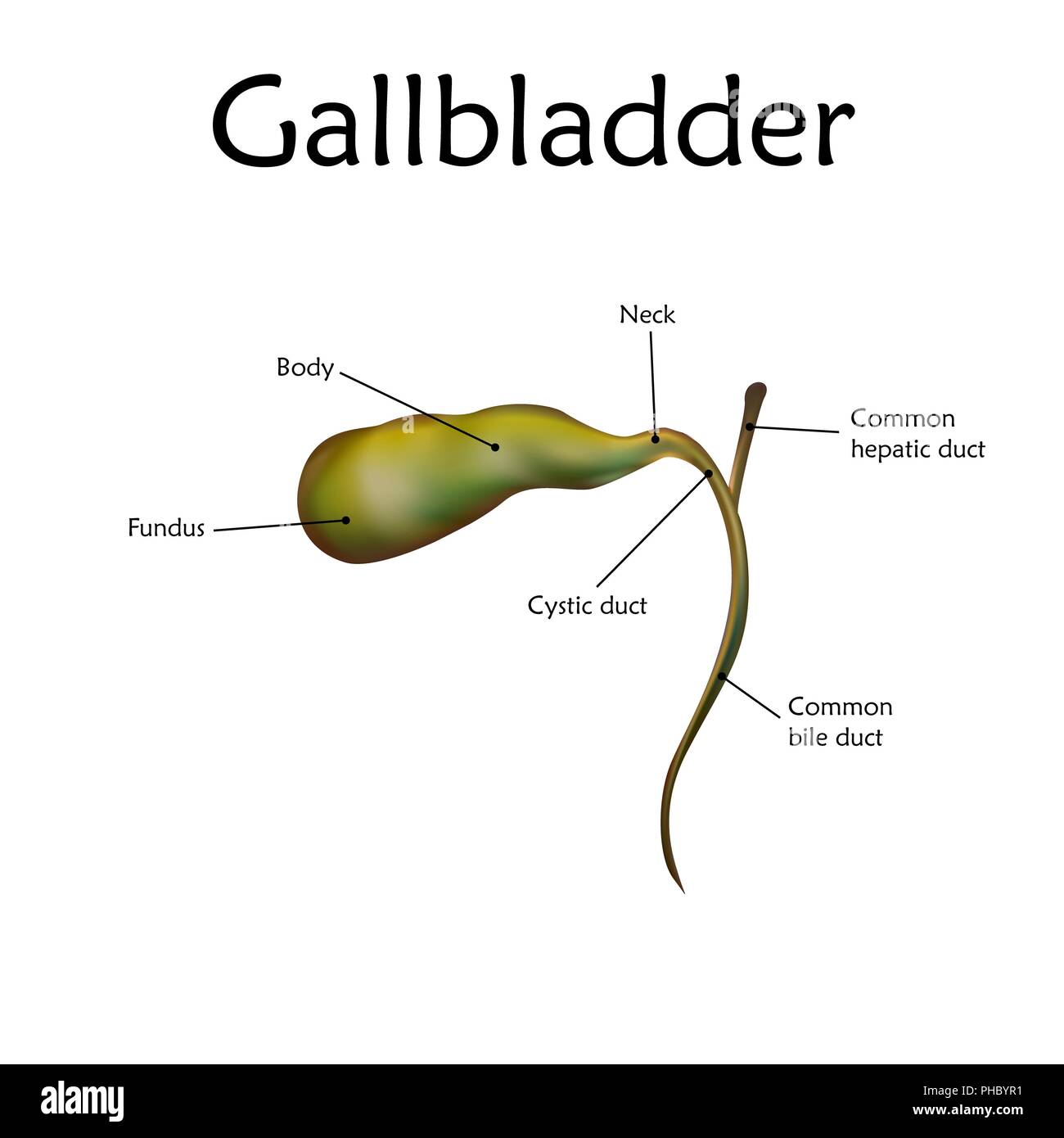
Gallbladder diagram hires stock photography and images Alamy

How to draw human Gallbladder step by step drawing tutorial YouTube

Cartoon human gallbladder anatomy Royalty Free Vector Image
The Gall Bladder Labeled Diagram Royalty Free Stock Vector 222549640
Gallbladder Definition, Anatomy, Parts, Function, Pictures eHealthStar

Gallbladder Drawing Free download on ClipArtMag
Gallbladder and Biliary Tract Anatomy Concise Medical Knowledge

Human gallbladder anatomy realistic illustration front view in detail
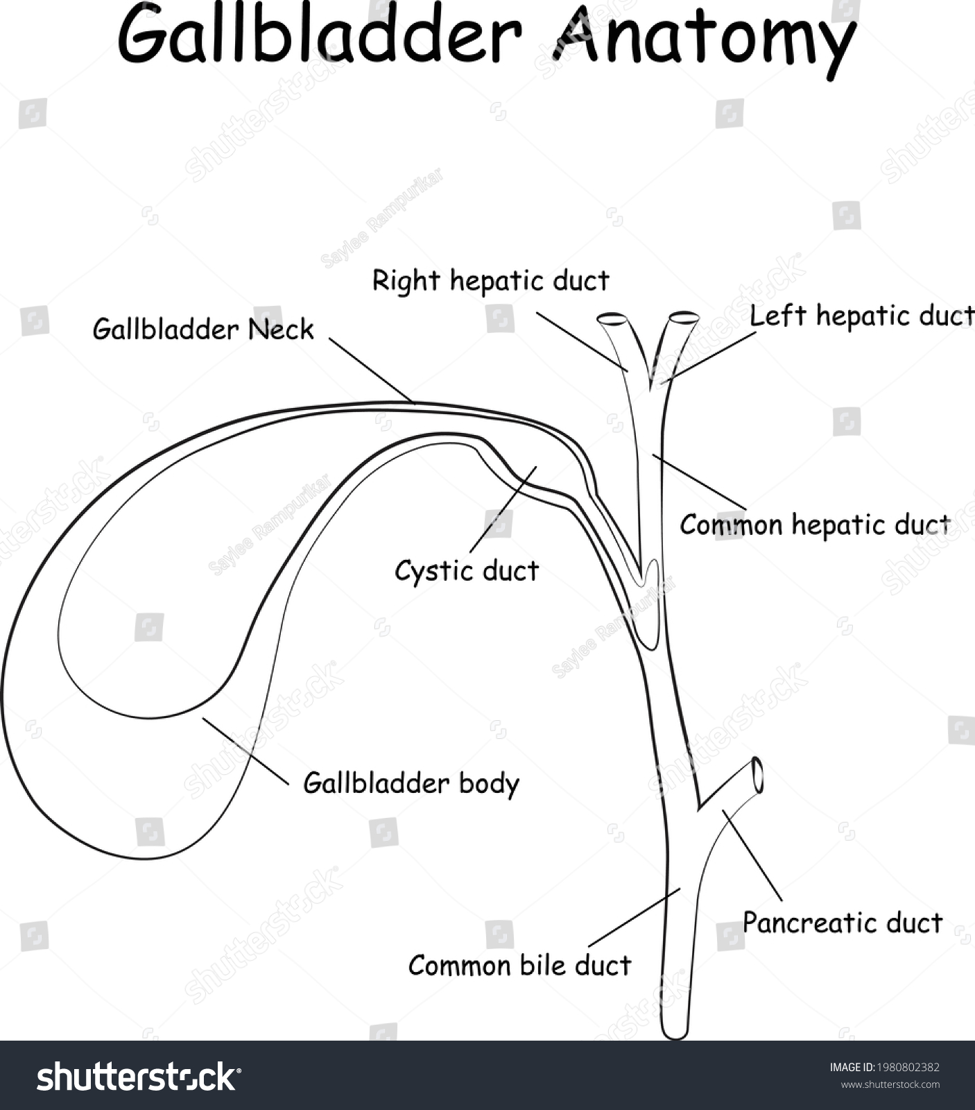
Gallbladder Anatomy Gallbladder Diagram Simple Line Stock Vector
Cartoon of Human Internal gallbladder anatomy Vector Image
Special Attention Will Also Be Paid To The Relevant Embryology, Histology And Adjacent Anatomical Structures.
Findings In This Randomized Clinical Trial Of 90 Participants, Both Gc And Ccrt.
Web Percutaneous Drainage Of The Gallbladder Involves Placing A Sterile Needle Into The Gallbladder To Aspirate (Draw Out) Bile.
A Gallbladder Scan Is A Type Of Nuclear.
Related Post: